Researchers from the University of California have programmed synthetic cells to mobilize nearby natural cells into complex structures. At first, individual cells self-organized into multi-layered structures resembling simple organisms or the tissues from the first stages of embryonic development. The technology could have a bright future in repairing damaged tissue or re-growing injured organs.
The fundamental question of developmental biology is how do complex biological structures emerge from a single fertilized egg. The deep understanding of these processes and the technology based on it could help thousands of people waiting for organ transplants. Today, scientists mostly place their hopes in growing and 3D-printing organs as means to combat the organ shortage. However, this technique is currently capable of producing only limited tissues and relies on the use of stem cells to generate mostly 2D structures like skin. In the study recently published in Science, researchers believe they might have found a way to make more organs viable for transplant. They used a new compound that mimics the DNA’s instructions for cells to turn into different tissues.
“People talk about 3D-printing organs, but that is really quite different from how biology builds tissues,” said study senior author Wendell Lim, chair of the department of cellular and molecular pharmacology at UCSF, for UCSF News. “Imagine if you had to build a human by meticulously placing every cell just where it needs to be and gluing it in place. It’s equally hard to imagine how you would print a complete organ, then make sure it was hooked up properly to the bloodstream and the rest of the body.”
During organ development, cells communicate one with another and make coordinated, collective decisions about how to structurally organize themselves. The UCSF research group used genetically modified cells to get other groups of individual cells to self-organize into multi-layered structures. Obtained structures were similar to simple organisms or early-stage tissues in human embryonic development.
“What is amazing about biology is that DNA allows all the instructions required to build an elephant to be packed within a tiny embryo,” said Lim. “It’s easy to get overwhelmed by the complexity of natural systems, so here we set out to understand the minimal set of rules for programming cells to self-assemble into multicellular structures.”
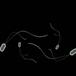
The new technique is made possible because of a customizable synthetic signaling molecule called synNotch, referring to a synthetic Notch receptor. The Delta-Notch signaling pathway is a highly conserved element in cell-cell communication across species. The synNotch cells, therefore, allow researchers to program other nearby niche cells with specific sets of instructions. For example, they engineered several groups of neighboring cells to produce “Velcro-like” adhesion molecules called cadherins along with fluorescent marker proteins. Specific directions were sent via the synNotch cells to induce neighboring cells to change color and self-organize into desired structures.
According to Lim, the synNotch technique is fundamentally different from other current techniques for tissue regeneration and growth. If researchers can find a way to program increasingly complex structures, then natural cellular development could take care of the rest. All that is left is to supply them with the blueprints.
“The beauty of self-organizing systems is that they are autonomous and compactly encoded,” Lim said. “You put in one or a few cells, and they grow and organize, taking care of the microscopic details themselves.”
Although the synNotch technique is in early stages and repairing tissue is still far away, the research group has set a good foundation. Surprisingly complex and important structures were already engineered. Some generated cells formed structures with a basic polarity which is the gateway to the development of functional complexity. These axes define the front-back, left-right and head-toe plans of individual organisms. By adding different types of cadherin adhesion molecules, researchers directed cellular assemblages to divide into “head” and “tail” sections, or to produce four distinct radial “arms.”
Watch an animation depicting the Notch intracellular signaling pathway:
Learn more about how signaling proteins are built from simple modules arranged in different ways, in Dr. Lim’s video below:
By Andreja Gregoric, MSc

")